History of anatomy
From Wikipedia, the free encyclopedia
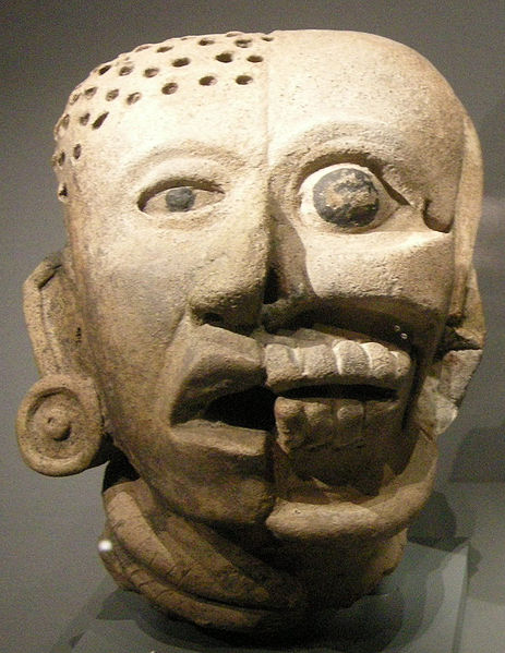
Maya bust
The development of anatomy as a science extends from the earliest examinations of sacrificial victims to the sophisticated analyses of the body performed by modern scientists. It has been characterized, over time, by a continually developing understanding of the functions of organs and structures in the body. The field of Human Anatomy has a prestigious history, and is considered to be the most prominent of the biological sciences of the 19th and early 20th centuries. Methods have also improved dramatically, advancing from examination of animals through dissection of cadavers to technologically complex techniques developed in the 20th century [1].
Anatomy is one of the cornerstones of a doctor’s medical education. Despite being a persistent portion of teaching from at least the renaissance, the format and the amount of information being taught has evolved and changed along with the demands of the profession. What is being taught today may differ in content significantly from the past but the methods used to teach this have not really changed that much. For example all the famous public dissections of the Middle Ages and early renaissance were in fact prosections. Prosection is the direction in which many current medical schools are heading in order to aid the teaching of anatomy and some argue that dissection is better. However looking at results of post graduate exams, medical schools (specifically Birmingham) that use prosection as opposed to dissection do very well in these examinations [2]. This would suggest that prosection can fit very well into the structure of modern medical training.
Ancient anatomy
Charaka is referred to as the Father of Anatomy.
[edit]
Egypt
The study of anatomy begins at least as early as 1600 BCE, the date of the Edwin Smith Surgical Papyrus. This treatise shows that the heart, its vessels, liver, spleen, kidneys, hypothalamus, uterus and bladder were recognized, and that the blood vessels were known to emanate from the heart. Other vessels are described, some carrying air, some mucus, and two to the right ear are said to carry the "breath of life", while two to the left ear the "breath of death". The Ebers papyrus (c. 1550 BCE) features a treatise on the heart. It notes that the heart is the center of the blood supply, with vessels attached for every member of the body. The Egyptians seem to have known little about the function of the kidneys and made the heart the meeting point of a number of vessels which carried all the fluids of the body – blood, tears, urine and sperm.[3]
[edit]
Greece
The earliest medical scientist of whose works any great part survives today is Hippocrates, a Greek physician active in the late 5th and early 4th centuries BCE (460 - 377 BCE). His work demonstrates a basic understanding of musculoskeletal structure, and the beginnings of understanding of the function of certain organs, such as the kidneys. Much of his work, however, and much of that of his students and followers later, relies on speculation rather than empirical observation of the body. One of the greatest achievements of Hippocrates was that he was the first to discover the tricuspid valve of the heart and its function which he documented in the treatise On the Heart in the Hippocratic Corpus. Later anatomists knew the function of the tricuspid valve after reading the Hippocratic Corpus.
In the 4th century BCE, Aristotle and several contemporaries produced a more empirically founded system, based animal dissection. Around this time, Praxagoras is credited as the first to identify the difference between arteries and veins, and the relations between organs are described more accurately than in previous works.
The first use of human cadavers for anatomical research occurred later in the 4th century BCE when Herophilos and Erasistratus gained permission to perform live dissections, or vivisection, on criminals in Alexandria under the auspices of the Ptolemaic dynasty. Herophilos in particular developed a body of anatomical knowledge much more informed by the actual structure of the human body than previous works had been.
[edit]
Galen
The final major anatomist of ancient times was Galen, active in the 2nd century. He compiled much of the knowledge obtained by previous writers, and furthered the inquiry into the function of organs by performing vivisection on animals. Due to a lack of readily available human specimens, discoveries through animal dissection were broadly applied to human anatomy as well. His collection of drawings, based mostly on dog anatomy, became the anatomy textbook for 1500 years. The original text is long gone, and his work was only known to the Renaissance doctors through the careful custody of Arabic medicine.
[edit]
Medieval anatomy
Main article: Islamic medicine - Anatomy and Physiology
After the fall of the Roman Empire, the study of anatomy became stagnant in Christian Europe but flourished in the medieval Islamic world, where Muslim physicians and Muslim scientists contributed heavily to medieval learning and culture. The Persian physician Avicenna (980-1037) absorbed the Galenic teachings on anatomy and expanded on them in The Canon of Medicine (1020s), which was very influential throughout the Islamic world and Christian Europe. The Canon remained the most authoritative book on anatomy in the Islamic world until Ibn al-Nafis in the 13th century, though the book continued to dominate European medical education for even longer until the 16th century.
The Arabian physician Ibn Zuhr (Avenzoar) (1091–1161) was the first physician known to have carried out human dissections and postmortem autopsy. He proved that the skin disease scabies was caused by a parasite, a discovery which upset the theory of humorism supported by Hippocrates and Galen. The removal of the parasite from the patient's body did not involve purging, bleeding, or any other traditional treatments associated with the four humours.[4] In the 12th century, Saladin's physician Ibn Jumay was also one of the first to undertake human dissections, and he made an explicit appeal for other physicians to do so as well. During a famine in Egypt in 1200, Abd-el-latif observed and examined a large number of skeletons, and he discovered that Galen was incorrect regarding the formation of the bones of the lower jaw and sacrum.[5]
[edit]
Ibn al-Nafis
The Arabian physician Ibn al-Nafis (1213–1288) was one of the earliest proponents of human dissection and postmortem autopsy, and in 1242, he was the first to describe the pulmonary circulation[6] and coronary circulation[7] of the blood, which form the basis of the circulatory system, for which he is considered the father of the theory of circulation.[8] Ibn al-Nafis also described the earliest concept of metabolism,[9] and developed new systems of anatomy and physiology to replace the Avicennian and Galenic doctrines, while discrediting many of their erroneous theories on the four humours, pulsation,[10] bones, muscles, intestines, sensory organs, bilious canals, esophagus, stomach, and the anatomy of almost every other part of the human body.[11]
[edit]
Early modern anatomy
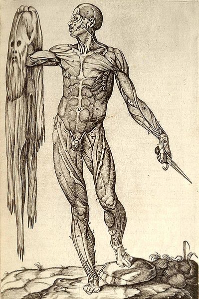
In this 1559 anatomical plate by Juan Valverde de Amusco, a figure holds a knife in one hand and his own skin in the other.
The works of Galen and Avicenna, especially The Canon of Medicine which incorporated the teachings of both, were translated into Latin, and the Canon remained the most authoritative text on anatomy in European medical education until the 16th century. The first major development in anatomy in Christian Europe, since the fall of Rome, occurred at Bologna in the 14th to 16th centuries, where a series of authors dissected cadavers and contributed to the accurate description of organs and the identification of their functions. Prominent among these anatomists were Mondino de Liuzzi and Alessandro Achillini.
The first challenges to the Galenic doctrine in Europe occurred in the 16th century. Thanks to the printing press, all over Europe a collective effort proceeded to circulate the works of Galen and Avicenna, and later publish criticisms on their works. Vesalius was the first to publish a treatise, De humani corporis fabrica, that challenged Galen "drawing for drawing" travelling all the way from Leuven[12] to Padua for permission to dissect victims from the gallows without fear of persecution. His drawings are triumphant descriptions of the, sometimes major, discrepancies between dogs and humans, showing superb drawing ability. Many later anatomists challenged Galen in their texts, though Galen reigned supreme for another century.
A succession of researchers proceeded to refine the body of anatomical knowledge, giving their names to a number of anatomical structures along the way. The 16th and 17th centuries also witnessed significant advances in the understanding of the circulatory system, as the purpose of valves in veins was identified, the left-to-right ventricle flow of blood through the circulatory system was described, and the hepatic veins were identified as a separate portion of the circulatory system. The lymphatic system was also identified as a separate system at this time.
[edit]
17th and 18th centuries
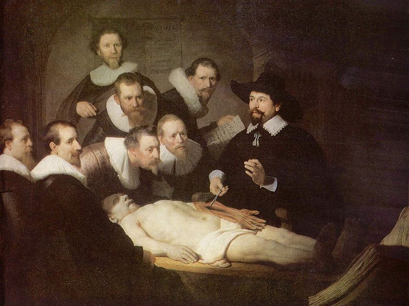
The Anatomy Lesson of Dr. Nicolaes Tulp, by Rembrandt, depicts an autopsy.
The study of anatomy flourished in the 17th and 18th centuries. The advent of the printing press facilitated the exchange of ideas. Because the study of anatomy concerned observation and drawings, the popularity of the anatomist was equal to the quality of his drawing talents, and one need not be an expert in Latin to take part. [2] Many famous artists studied anatomy, attended dissections, and published drawings for money, from Michelangelo to Rembrandt. For the first time, prominent universities could teach something about anatomy through drawings, rather than relying on knowledge of Latin. Contrary to popular belief,the church neither objected to nor obstructed anatomical research despite its antagonism towards other scientific practices.[13]. The increase in demand for cadavers, though, led to rumors about anatomy murder.
Only certified anatomists were allowed to perform dissections, and sometimes then only yearly. These dissections were sponsored by the city councilors and often charged an admission fee, rather like a circus act for scholars. Many European cities, such as Amsterdam, London, Copenhagen, Padua, and Paris, all had Royal anatomists (or some such office) tied to local government. Indeed, Nicolaes Tulp was Mayor of Amsterdam for three terms. Though it was a risky business to perform dissections, and unpredictable depending on the availability of fresh bodies, attending dissections was perfectly legal. Many anatomy students traveled around Europe from dissection to dissection during the course of their study - they had to go where a fresh body was available (e.g. after a hanging) because before refrigeration, a body would decay rapidly and become unsuitable for examination.
Many Europeans interested in the study of anatomy traveled to Italy, then the center of anatomy. Only in Italy could certain important research methods be used, such as dissections on women. M. R. Columbus and Gabriele Falloppio were pupils of Vesalius, the 16th century anatomist. Columbus, as his immediate successor in Padua, and afterwards professor at Rome, distinguished himself by rectifying and improving the anatomy of the bones, by giving correct accounts of the shape and cavities of the heart, of the pulmonary artery and aorta and their valves, and tracing the course of the blood from the right to the left side of the heart, by a good description of the brain and its vessels, and by correct understanding of the internal ear, and the first good account of the ventricles of the larynx. Osteology at nearly the same time found an assiduous cultivator in Giovanni Filippo Ingrassias.
[edit]
19th century anatomy
Further information: History of anatomy in the 19th century
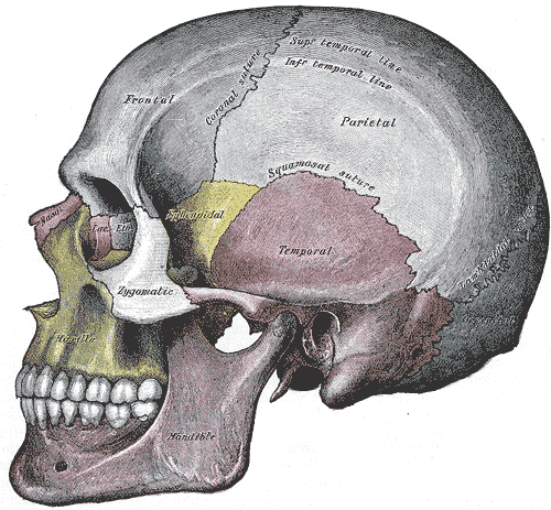
A plate of the skull from the 1918 edition of Gray's Anatomy
During the 19th century, anatomists largely finalised and systematised the descriptive human anatomy of the previous century. The discipline also progressed to establish growing sources of knowledge in histology and developmental biology, not only of humans but also of animals. Extensive research was conducted in more areas of anatomy. Great Britain was particularly important in this research.[citation needed] Demand for cadavers grew so great there that body-snatching and even anatomy murder came into use as a means of obtaining them.[14] In response, the English Parliament passed the Anatomy Act 1832, which finally provided for an adequate and legitimate supply of corpses by allowing dissection of destitutes. The relaxed restrictions on dissection provided a suitable environment for Gray's Anatomy, a text that was a collective effort and became widely popular. Now seen as unwieldy, Gray's Anatomy was born out of a need to create a single volume on anatomy for the traveling doctor.
The shift from the largely public displays of dissection in anatomy theatres to dissections carried out in classrooms meant that there was a drastic change in who could observe a dissection. Females for example, who at this time were not allowed to attend medical school, could broaden their knowledge by attending the anatomy theatres. So the shift from prosection to dissection meant a reduction in the number of people that could benefit from a single cadaver. At this point as well tighter regulation of the medical profession and donations of bodies resulted in various implications for carrying out dissections. Private medical schools which offered summer schools and various other courses involving cadaveric dissection allowed one route into gaining membership to the Royal College of Surgeons. However from 1822 the Royal College of surgeons would no longer accept these qualifications, this as result would see these largely unregulated schools begin to close [15]. Not only as a result of this, but the Anatomy Act 1832 made it much harder (more bureaucracy) to obtain bodies for dissection. The act resulted in only the large teaching hospitals feasibly being able to continue teaching anatomy courses due to agreements with patients that if they donated their body they would receive free treatment. So towards the end of 19th century anatomy courses had been largely professionalised at established medical schools and public dissection was no longer common place.
Another source of anatomy teaching began with the foundation of many medical schools (particularly within the provincial medical schools) and the medical museums found within them. A large portion of training occurred within these up until and for some time after the Second World War. The medical museum was very important and a lot of effort was put into creating something impressive. This was particularly so in provincial medical schools which were just being established that needed credibility not only from other medical schools (namely Oxford and the London teaching hospitals) but also from the public. The museums were not only for students but also members of the public paid to see the exhibits within the museum. This brought not only much needed income but prestige as well [16]. The more exhibits within the museum the more established the medical school appeared to be (at least to the public). Significant amounts of teaching occurred in the museum as well with students claiming they learnt far more in the museum than they ever did in the lecture theatre. The decline of the museums within medical schools was largely due to the demand in floor space for teaching and new disciplines and less importantly the great improvements in photography and colour texts. For example the museum at Birmingham Medical School is now a computer cluster and teaching rooms, the only remains of the museum are the preserved specimens decorating the walls around the computer cluster.
[edit]
Modern anatomy
Anatomical research in the past hundred years has taken advantage of technological developments and growing understanding of sciences such as evolutionary and molecular biology to create a thorough understanding of the body's organs and structures. Disciplines such as endocrinology have explained the purpose of glands that anatomists previously could not explain; medical devices such as MRI machines and CAT scanners have enabled researchers to study the organs of living people or of dead ones. Progress today in anatomy is centered in the development, evolution, and function of anatomical features, as the macroscopic aspects of human anatomy have been largely catalogued. The subfield of non-human anatomy is particularly active as modern anatomists seek to understand basic organizing principles of anatomy through the use of advanced techniques ranging from finite element analysis to molecular biology.
With increasing demands on the healthcare system and what could be deemed chronic under-training of doctors (numbers of doctors per capita compared to other industrialised countries) during the latter half of the 20th century, medical schools are now facing massive pressure to train as many doctors as possible. This has meant in recent years cohort sizes have doubled and more in size, in order to try and meet the demand. This has resulted in increased pressure of the facilities at all medical schools in the country. Anatomy is one department in particular that has had to evolve to accommodate the number of students. At Birmingham dissection was once essential to the teaching of anatomy but since the end of the 1980s the medical school has adopted prosection over dissection. At the time new directives from the General Medical Council (GMC) on the direction medical education was the major factor according the current head of anatomy. There are also many other reasons why prosection maybe favoured (discussed below). It has probably now become near impossible to restart dissection at Birmingham even if one wanted to. This is due to the fact that current prosection uses a very similar number of cadavers as dissection previously did. If dissection was to be brought back the number of cadavers would be very large due the current cohort size. To increase provision of prosection the medical school is currently investing in the region of £800,000-900,000 on a new prosectorium. This will allow up to about 40 students to observe prosected material in any one session. The vast amount of money required just to increase the amount of prosection demonstrates that it is no longer possible to carry out dissection at Birmingham (and is the case for many other universities). Prosection makes more efficient use of a cadaver when compared to dissection. A single cadaver when dissecting would be used by up to 5 students whereas prosection allows if necessary and entire cohort to observe the prosected cadaver. Prosection also allows students to observe more than one cadaver whereas in dissection you would tend to just use a single one. Logistically prosection allows more flexibility than dissection as there is no commitment to provide a cadaver per a certain number of students, this in fact create opportunities for cadavers to be used, for example at Birmingham, for Special Study Modules (SSMs) and postgraduate teaching.
Also there are many more aids to teaching anatomy then merely the prosectorium; improvements over the last century in colour images and photographs means that an anatomy text is no longer an aid to dissection but rather a central material to learn from. Plastic models are also regularly used in anatomy teaching sessions and they offer a good substitute to the real thing. One argument against plastic models is that they may provide a false sense of conformity in the human body; there is no doubt quite a difference between a plastic model and a prosected cadaver. Use of living models for anatomy demonstration is once again becoming popular within teaching of anatomy. Anatomy is dynamic, for example the anatomy of the musculoskeletal system is by definition the anatomy of movement. So to provide an example of this to the audience (students) and be able to demonstrate the possible movements is beneficial. Surface landmarks that can be palpated on another individual also provide practice for future clinical situations. It is possible to do this on oneself and a good example of this being implemented is Integrated Biology at the University of Berkeley; students are encouraged to “introspect” [17] on themselves and link what they are being taught to their own body. This may seem like a relatively obvious idea but to formally link it into teaching of anatomy should aid memory recall [15].
Donations of bodies have also declined in recent years with a marked decline of public confidence in the medical profession. With scandals such as Alder hay and Bristol, people are less confident that their wishes on what will happen to their body will be carried out, so instead have not donated to medical science when in the past they may have [18]. The resultant legislation from these scandals (namely the Human Tissue Act 2004) has tightened up the availability of resources to anatomy departments. Another factor facing body donations is the problems arising from the outbreaks of Bovine Spongiform Encephalitis (BSE) in the late 80s and early 90s and the restrictions of handling of brain tissue that resulted from this. The exact pathology of the human form, variant Creutzfeldt–Jakob disease (vCJD) has meant that patients donating their body who suffered from Alzheimer’s or dementia and of course vCJD means their brains cannot be handled. As the method of transmission of these diseases and the link between them (i.e. is Alzheimer’s vCJD and vice versa) is not fully understood these precautions have to be taken [19]. Very symptomatic patients are also not normally accepted for cadavers [15]. However this means that students are more limited on what they can dissect within the head, this is particularly a problem in medical schools where dissection is still carried out. It is less of a problem where prosection is carried out as the specimen will have already been dissected.
[edit]
Conclusion
Anatomy teaching has changed considerably over the last 1000 years though it is still very much at the heart of the philosophy of western medicine. Western medicine seeks to find a cause to all disease and attempt to cure it; very much cause and effect. Without a good understanding of the arrangement of the human body then this becomes somewhat challenging. Western medicine is in fact taking a more holistic approach today, with the psychosocial biomedical model of disease. However most practicing doctors if it was proven that there was a biological cause to all the various idiopathic diseases then they would readily adapt their thoughts and treatments accordingly. Anatomy is often regarded as being little left to discover, in that we know what and where most of the body is and does, but there is still many mysteries left to work out. Public awareness of anatomy cannot be detrimental if it sparks interest in the discipline. The recent controversies with Gunther von Hagens and public displays of dissection may divide opinions on what is ethical (even the legality of a public dissection) [20] but this surely at least gets people thinking about how doctors learn anatomy and why in some it inspires them to pursue a career. The future of dissection may be uncertain and indeed if pressure on cadavers continues even the few medical schools that continue to do dissection may have to halt. This hopefully however will not reduce the number of people able to benefit from a single cadaver if current prosection methods become the prevalent method of demonstrating gross anatomy.
Anatomy
From Wikipedia, the free encyclopedia
For other uses, see anatomy (disambiguation). Wikiversity has learning materials about Topic:Anatomy
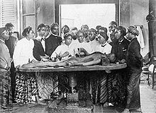
Anatomy lesson carried out in Java, Dutch East Indies, date unknown.
Anatomy (from the Greek ἀνατομία anatomia, from ἀνατέμνειν ana: separate, apart from, and temnein, to cut up, cut open) is a branch of biology and medicine that is the consideration of the structure of living things. It is a general term that includes human anatomy, animal anatomy (zootomy) and plant anatomy (phytotomy). In some of its facets anatomy is closely related to embryology, comparative anatomy and comparative embryology,[1] through common roots in evolution.
Anatomy is subdivided into gross anatomy (or macroscopic anatomy) and microscopic anatomy.[1] Gross anatomy (also called topographical anatomy, regional anatomy, or anthropotomy) is the study of anatomical structures that can be seen by unaided vision with the naked eye.[1] Microscopic anatomy is the study of minute anatomical structures assisted with microscopes, which includes histology (the study of the organization of tissues),[1] and cytology (the study of cells).
The history of anatomy has been characterized, over time, by a continually developing understanding of the functions of organs and structures in the body. Methods have also improved dramatically, advancing from examination of animals through dissection of cadavers (dead human bodies) to technologically complex techniques developed in the 20th century including X-ray, ultrasound, and MRI imaging.
Anatomy should not be confused with anatomical pathology (also called morbid anatomy or histopathology), which is the study of the gross and microscopic appearances of diseased organs.
Superficial anatomy
Superficial anatomy or surface anatomy is important in anatomy being the study of anatomical landmarks that can be readily seen from the contours or the surface of the body.[1] With knowledge of superficial anatomy, physicians or veterinary surgeons gauge the position and anatomy of the associated deeper structures. Superficial is a directional term that indicates one structure is located more externally than another, or closer to the surface of the body.
[edit]
Human anatomy
Main article: Human anatomy
Para-sagittal MRI scan of the head
An X-ray of a human chest.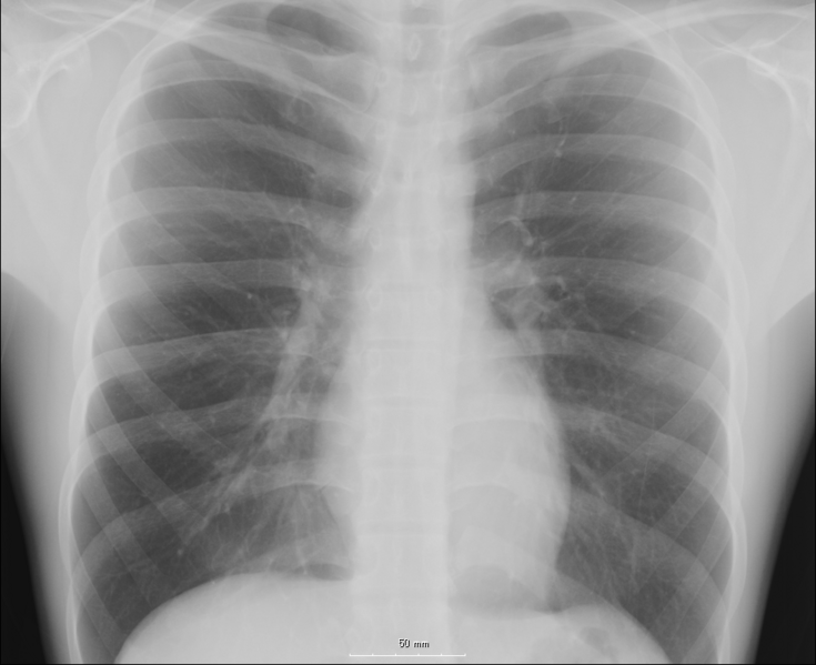
Human heart and lungs, from an older edition of Gray's Anatomy.
Human anatomy, including gross human anatomy and histology, is primarily the scientific study of the morphology of the adult human body.[1]
Generally, students of certain biological sciences, paramedics, prosthetists and orthotists, physiotherapists, occupational therapy, nurses, and medical students learn gross anatomy and microscopic anatomy from anatomical models, skeletons, textbooks, diagrams, photographs, lectures and tutorials. The study of microscopic anatomy (or histology) can be aided by practical experience examining histological preparations (or slides) under a microscope; and in addition, medical students generally also learn gross anatomy with practical experience of dissection and inspection of cadavers (dead human bodies).
Human anatomy, physiology and biochemistry are complementary basic medical sciences, which are generally taught to medical students in their first year at medical school. Human anatomy can be taught regionally or systemically;[1] that is, respectively, studying anatomy by bodily regions such as the head and chest, or studying by specific systems, such as the nervous or respiratory systems. The major anatomy textbook, Gray's Anatomy, has recently been reorganized from a systems format to a regional format,[2][3] in line with modern teaching methods. A thorough working knowledge of anatomy is required by all medical doctors, especially surgeons, and doctors working in some diagnostic specialities, such as histopathology and radiology.
Academic human anatomists are usually employed by universities, medical schools or teaching hospitals. They are often involved in teaching anatomy, and research into certain systems, organs, tissues or cells.
[edit]
Other branches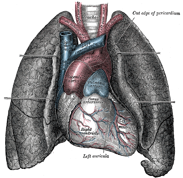
Comparative anatomy relates to the comparison of anatomical structures (both gross and microscopic) in different animals.[1]
Anthropological anatomy or physical anthropology relates to the comparison of the anatomy of different races of humans.
Artistic anatomy relates to anatomic studies for artistic reasons.
Branches of biology

Botany
From Wikipedia, the free encyclopedia
"Plant biology" redirects here. For the journal, see Functional Plant Biology.
For other uses, see Botany (disambiguation) and Botanic (disambiguation).
Pinguicula grandiflora commonly known as a Butterwort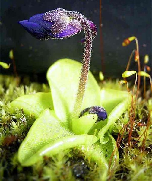
Botany, plant science(s), phytology, or plant biology is a branch of biology that involves the scientific study of plant life. Botany covers a wide range of scientific disciplines concerned with the study of plants, algae and fungi, including structure, growth, reproduction, metabolism, development, diseases, chemical properties, and evolutionary relationships among taxonomic groups. Botany began with early human efforts to identify edible, medicinal and poisonous plants, making it one of the oldest sciences. Today botanists study over 550,000 species of living organisms.
Scope and importance of botany
Hibiscus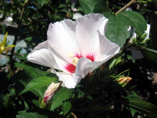
As with other life forms in biology, plant life can be studied from different perspectives, from the molecular, genetic and biochemical level through organelles, cells, tissues, organs, individuals, plant populations, and communities of plants. At each of these levels a botanist might be concerned with the classification (taxonomy), structure (anatomy and morphology), or function (physiology) of plant life.
Historically all living things were grouped as animals or plants,[1] and botany covered all organisms not considered animals. Some organisms once included in the field of botany are no longer considered to belong to the plant kingdom – these include fungi (studied in mycology), lichens (lichenology), bacteria (bacteriology), viruses (virology) and single-celled algae, which are now grouped as part of the Protista. However, attention is still given to these groups by botanists, and fungi, lichens, bacteria and photosynthetic protists are usually covered in introductory botany courses.
The study of plants is vital because they are a fundamental part of life on Earth, which generates the oxygen, food, fibres, fuel and medicine that allow humans and other life forms to exist. Through photosynthesis, plants absorb carbon dioxide, a greenhouse gas that in large amounts can affect global climate. Additionally, they prevent soil erosion and are influential in the water cycle. A good understanding of plants is crucial to the future of human societies as it allows us to:
Produce food to feed an expanding population
Understand fundamental life processes
Produce medicine and materials to treat diseases and other ailments
Understand environmental changes more clearly
Paleobotanists study ancient plants in the fossil record. It is believed that early in the Earth's history, the evolution of photosynthetic plants altered the global atmosphere of the earth, changing the ancient atmosphere by oxidation.
[edit]
Human nutrition
Nearly all the food we eat comes (directly and indirectly) from plants like this American long grain rice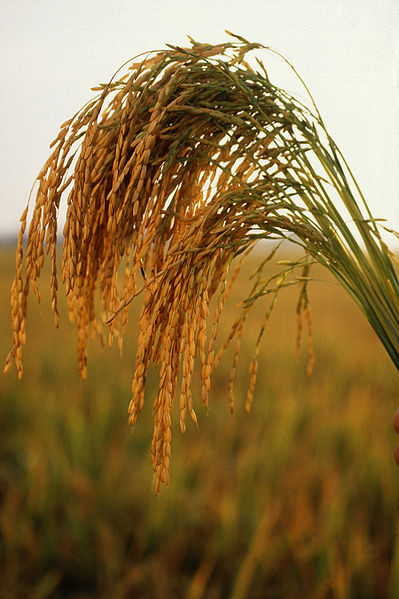
Virtually all foods eaten come from plants, either directly from staple foods and other fruit and vegetables, or indirectly through livestock or other animals, which rely on plants for their nutrition. Plants are the fundamental base of nearly all food chains because they use the energy from the sun and nutrients from the soil and atmosphere, converting them into a form that can be consumed and utilized by animals; this is what ecologists call the first trophic level. Botanists also study how plants produce food we can eat and how to increase yields and therefore their work is important in mankind's ability to feed the world and provide food security for future generations, for example, through plant breeding. Botanists also study weeds, plants which are considered to be a nuisance in a particular location. Weeds are a considerable problem in agriculture, and botany provides some of the basic science used to understand how to minimize 'weed' impact in agriculture and native ecosystems. Ethnobotany is the study of the relationships between plants and people.
[edit]
Fundamental life processes
Plants are convenient organisms in which fundamental life processes (like cell division and protein synthesis) can be studied, without the ethical dilemmas of studying animals or humans. The genetic laws of inheritance were discovered in this way by Gregor Mendel, who was studying the way pea shape is inherited. What Mendel learned from studying plants has had far reaching benefits outside of botany. Additionally, Barbara McClintock discovered 'jumping genes' by studying maize. These are a few examples that demonstrate how botanical research has an ongoing relevance to the understanding of fundamental biological processes.
[edit]
Medicine and materials
Many medicinal and recreational drugs, like tetrahydrocannabinol, caffeine, and nicotine come directly from the plant kingdom. Others are simple derivatives of botanical natural products; for example, aspirin is based on the pain killer salicylic acid which originally came from the bark of willow trees. As well, the narcotic analgesics such as morphine are derived from the opium poppy.[2] There may be many novel cures for diseases provided by plants, waiting to be discovered. Popular stimulants like coffee, chocolate, tobacco, and tea also come from plants. Most alcoholic beverages come from fermenting plants such as barley (beer), rice (sake) and grapes (wine).
Plants also provide us with many natural materials, such as hemp, cotton, wood, paper, linen, vegetable oils, some types of rope, and rubber. The production of silk would not be possible without the cultivation of the mulberry plant. Sugarcane, rapeseed, soy and other plants with a highly fermentable sugar or oil content have recently been put to use as sources of biofuels, which are important alternatives to fossil fuels (see biodiesel).
[edit]
Environmental changes
Plants can also help us understand changes in on our environment in many ways.
Understanding habitat destruction and species extinction is dependent on an accurate and complete catalog of plant systematics and taxonomy.
Plant responses to ultraviolet radiation can help us monitor problems like ozone depletion.
Analyzing pollen deposited by plants thousands or millions of years ago can help scientists to reconstruct past climates and predict future ones, an essential part of climate change research.
Recording and analyzing the timing of plant life cycles are important parts of phenology used in climate-change research.
Lichens, which are sensitive to atmospheric conditions, have been extensively used as pollution indicators.
In many different ways, plants can act a little like the 'miners' canary', an early warning system alerting us to important changes in our environment. In addition to these practical and scientific reasons, plants are extremely valuable as recreation for millions of people who enjoy gardening, horticultural and culinary uses of plants every day.
[edit]
Etymology
From Greek βοτάνη = "pasture, grass, fodder", perhaps via the idea of a livestock keeper needing to know which plants are safe for livestock to eat.
[edit]
History
Main article: History of botany
The traditional tools of a botanist
[edit]
Early botany
Ancient India

Early examples of plant taxonomy occur in the Rigveda, that divides plants into Vṛska (tree), Osadhi (herbs useful to humans) and Virudha (creepers), which are then further subdivided. The Atharvaveda divides plants into eight classes, Visakha (spreading branches), Manjari (leaves with long clusters), Sthambini (bushy plants), Prastanavati (which expands); Ekasṛnga (those with monopodial growth), Pratanavati (creeping plants), Amsumati (with many stalks), and Kandini (plants with knotty joints). The Taittiriya Samhita classifies the plant kingdom into vṛksa, vana and druma (trees), visakha (shrubs with spreading branches), sasa (herbs), amsumali (a spreading or deliquescent plant), vratati (climber), stambini (bushy plant), pratanavati (creeper), and alasala (those spreading on the ground).
Manusmriti – Law book of Hindus – proposed a classification of plants in eight major categories. Charaka Samhitā and Sushruta Samhita and the Vaisesikas also present an elaborate taxonomy.
Parashara, the author of Vṛksayurveda (the science of life of trees), classifies plants into Dvimatrka (Dicotyledons) and Ekamatrka (Monocotyledons). These are further classified into Samiganiya (Fabaceae), Puplikagalniya (Rutaceae), Svastikaganiya (Cruciferae), Tripuspaganiya (Cucurbitaceae), Mallikaganiya (Apocynaceae), and Kurcapuspaganiya (Asteraceae).[3]
Important medieval Indian works of plant physiology include the Prthviniraparyam of Udayana, Nyayavindutika of Dharmottara, Saddarsana-samuccaya of Gunaratna, and Upaskara of Sankaramisra.[3]
Ancient Iranic people
The knowledge of medical plants and botany was considered as secret and holy by the ancient Iranic people. There is evidence of such practices in the documents that have survived from the ancient Zoroastrian writings. The practice and use of botany for medical purposes as well as various Iranic cousins and traditions is still common to this day amongst the Iranic people of the Central Asia, Near East and Europe.
Ancient China
In ancient China, the recorded listing of different plants and herb concoctions for pharmaceutical purposes spans back to at least the Warring States (481 BC-221 BC). Many Chinese writers over the centuries contributed to the written knowledge of herbal pharmaceutics. There was the Han Dynasty (202 BC-220 AD) written work of the Huangdi Neijing and the famous pharmacologist Zhang Zhongjing of the 2nd century. There was also the 11th century scientists and statesmen Su Song and Shen Kuo, who compiled treatises on herbal medicine and included the use of mineralogy.
Greco-Roman world
Among the earliest of botanical works in Europe, written around 300 B.C., are two large treatises by Theophrastus: On the History of Plants (Historia Plantarum) and On the Causes of Plants. Together these books constitute the most important contribution to botanical science during antiquity and on into the Middle Ages. Aristotle also wrote about plants. One theory about plants that Greco-Romans came up with about plants was that they ate soil for nutrients.[4]
The Roman medical writer Pedanius Dioscorides (ca.40-90) provides important evidence on Greek and Roman knowledge of medicinal plants. Dioscorides is famous for writing a five volume book in his native Greek Περί ύλης ιατρικής (De Materia Medica - in the Latin translation) that is one of the most influential herbal books in history. In fact, it remained in use until about CE 1600.[5] Approximately 1300-1400 different plant species were known under Roman reign.[6]
[edit]
Medieval botany
Main article: Muslim Agricultural Revolution
The earliest known work from the Muslim world dedicated to the study of agriculture was Ibn Wahshiyya's Nabatean Agriculture, which also dealt with the related field of botany and was also an early cookbook.
The Kurdish biologist Abū Ḥanīfa Dīnawarī (828-896) is considered the founder of Arabic botany for his Book of Plants, in which he described at least 637 plants and discussed plant development from germination to death, describing the phases of plant growth and the production of flowers and fruit.[7]
Theophrastus’s Historia Plantarum served as a reference point in botany for many centuries, and was further developed around 1200 A.D. by Giovanni Bodeo da Stapelio, who added a commentarius and drawings: see Historia Plantarum —Selected pages of a 17th century edition of the 1200 A.D. version (in Italian).
Ibn Bassal is known for his famous work named The Classification of Soils. Al-Asma'i was the earliest known Arab biologist, botanist and zoologist. al-Masihi was the first to recognize the science of Botany.[citation needed]
In the early 13th century, the Andalusian-Arabian biologist Abu al-Abbas al-Nabati developed an early scientific method for botany, introducing empirical and experimental techniques in the testing, description and identification of numerous materia medica, and separating unverified reports from those supported by actual tests and observations.[8][verification needed] His student Ibn al-Baitar (d. 1248) wrote a pharmaceutical encyclopedia describing 1,400 plants, foods, and drugs, 300 of which were his own original discoveries. A Latin translation of his work was useful to European biologists and pharmacists in the 18th and 19th centuries.[9][verification needed]
[edit]
Early modern botany
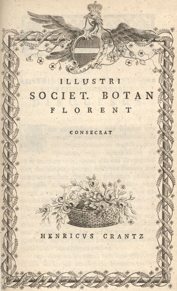
Crantz's Classis cruciformium..., 1769
German physician Leonhart Fuchs (1501–1566) was one of the three founding fathers of botany, along with Otto Brunfels (1489- 1534) and Hieronymus Bock (1498–1554) (also called Hieronymus Tragus).[10]
Valerius Cordus (1515–1554) authored one of the greatest pharmacopoeias and one of the most celebrated herbals in history, Dispensatorium (1546).[11] As early as the 16th century, the Italian Ulisse Aldrovandi was scientifically researching plants. In 1665, using an early microscope, Robert Hooke discovered cells in cork, and a short time later in living plant tissue. The Germans Jacob Theodor Klein and Leonhart Fuchs, the Swiss Conrad von Gesner, and the British author Nicholas Culpeper published herbals that gave information on the medicinal uses of plants.
During the 18th century systems of classification became deliberately artificial and served only for the purpose of identification. These classifications are comparable to diagnostic keys, where taxa are artificially grouped in pairs by few, easily recognisable characters. The sequence of the taxa in keys is often totally unrelated to their natural or phyletic groupings. In the 18th century an increasing number of new plants had arrived in Europe, from newly discovered countries and the European colonies worldwide, and a larger amount of plants became available for study.
In 1754 Carl von Linné (Carl Linnaeus) divided the plant Kingdom into 25 classes. One, the Cryptogamia, included all the plants with concealed reproductive parts (algae, fungi, mosses and liverworts and ferns).[12]
The increased knowledge on anatomy, morphology and life cycles, lead to the realization that there were more natural affinities between plants, than the sexual system of Linnaeus indicated. Adanson (1763), de Jussieu (1789), and Candolle (1819) all proposed various alternative natural systems that were widely followed. The ideas of natural selection as a mechanism for evolution required adaptations to the Candollean system, which started the studies on evolutionary relationships and phylogenetic classifications of plants.
[edit]
Modern botany
A considerable amount of new knowledge today is being generated from studying model plants like Arabidopsis thaliana. This weedy species in the mustard family was one of the first plants to have its genome sequenced. The sequencing of the rice (Oryza sativa) genome, its relatively small genome, and a large international research community have made rice an important cereal/grass/monocot model.[13] Another grass species, Brachypodium distachyon is also emerging as an experimental model for understanding the genetic, cellular and molecular biology of temperate grasses. Other commercially important staple foods like wheat, maize, barley, rye, pearl millet and soybean are also having their genomes sequenced. Some of these are challenging to sequence because they have more than two haploid (n) sets of chromosomes, a condition known as polyploidy, common in the plant kingdom. Chlamydomonas reinhardtii (a single-celled, green alga) is another plant model organism that has been extensively studied and provided important insights into cell biology.
In 1998 the Angiosperm Phylogeny Group published a phylogeny of flowering plants based on an analysis of DNA sequences from most families of flowering plants. As a result of this work, major questions such as which families represent the earliest branches in the genealogy of angiosperms are now understood. Investigating how plant species are related to each other allows botanists to better understand the process of evolution in plants.
[edit]
Subdisciplines of botanyAgronomy — Application of plant science to crop production
Bryology — Mosses, liverworts, and hornworts
Economic botany — Study of plants of economic use or value
Ethnobotany — Relationship between humans and plants
Forestry — Forest management and related studies
Horticulture — Cultivated plants
Lichenology — The study of lichens
Paleobotany — Fossil plants
Palynology — Pollen and spores Phycology — Algae
Phytochemistry — Plant secondary chemistry and chemical processes
Phytopathology — Plant diseases
Plant anatomy — Cell and tissue structure
Plant ecology — Role of plants in the environment
Plant genetics — Genetic inheritance in plants
Plant morphology — Structure and life cycles
Plant physiology — Life functions of plants
Plant systematics — Classification and naming of plants
[edit]
Notable botanists
Further information: List of botanists
Isabella Abbott, world's leading expert on Hawaiian seaweeds; discovered over 200 species.
Ibn al-Baitar (d. 1248), Andalusian-Arab scientist, botanist, pharmacist, physician, and author of one of the largest botanical encyclopedias.
L.J.F. Brimble (1904–1965), English botanist and editor of Nature magazine
Abu al-Abbas al-Nabati (c. 1200), Andalusian-Arab botanist and agricultural scientist, and a pioneer in experimental botany.
Aimé Bonpland (1773–1858), French explorer and botanist, who accompanied Alexander von Humboldt during five years of travel in Latin America.
Luther Burbank (1849–1926), American botanist, horticulturist, and a pioneer in agricultural science.
Augustin Pyramus de Candolle (1778–1841), He originated the idea of "Nature's war", which influenced Charles Darwin.
Abū Ḥanīfa Dīnawarī (828-896), Persian botanist, historian, geographer, astronomer, mathematician, and founder of Arabic botany.
David Douglas (1799–1834), Scottish botanical explorer of North America and China, who imported many ornamental plants into Europe.
Joseph Dalton Hooker (1817–1911), English botanist and explorer. Second winner of Darwin Medal.
Pedanius Dioscorides (ca. 40-90 AD), physician, pharmacologist, toxicologist and botanist, author of Περὶ ὕλης ἰατρικής (Latin: De Materia Medica, English: "Regarding Medical Matters")
Thomas Henry Huxley (1825–1895), English biologist, known as "Darwin's Bulldog" for his advocacy of Charles Darwin's theory of evolution. Third winner of Darwin Medal.
Carl Linnaeus (1707–1778), Swedish botanist, physician and zoologist who laid the foundations for the modern scheme of Binomial nomenclature. He is known as the father of modern taxonomy, and is also considered one of the fathers of modern ecology.
Gregor Johann Mendel (1822–1884), Augustinian priest and scientist, and is often called the father of genetics for his study of the inheritance of traits in pea plants.
Charles Sprague Sargent (1841–1927), American botanist, the first director of the Arnold Arboretum at Harvard University.
Carlos Muñoz Pizarro (1913–1976), Chilean botanist, known for his studies of the Chilean flora, and its conservation.
Richard Spruce (1817–1893), English botanist and explorer who carried out a detailed study of the Amazon flora.
Agustín Stahl (1842–1917), conducted investigations and experiments in the fields of ethnology, and zoology in the Caribbean region.
George Ledyard Stebbins, Jr. (1906–2000), widely regarded as one of the leading evolutionary biologists of the 20th century, developed a comprehensive synthesis of plant evolution incorporating genetics.
Theophrastus (c. 371 – c. 287 BC), father of botany, established botanical science through his lecture notes, Enquiry into Plants.
Leonardo da Vinci (1452–1519), Italian polymath; a scientist, mathematician, engineer, inventor, anatomist, painter, sculptor, architect, botanist, musician and writer.
 THANK YOU FOR VISITING OUR SITE......................


